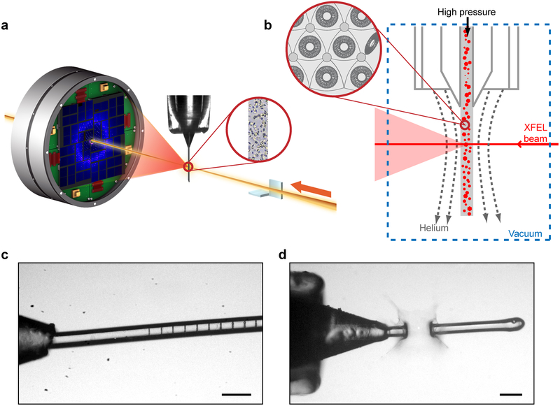
Figure 3. SFX-LCP process.
(a) Schematic of an LCP-SFX data collection setup. XFEL beam is focused to a diameter of ~1 μm by a pair of KB mirrors on a stream of LCP delivering micrometer-sized crystals intersecting the beam in random orientations. Diffraction patterns are collected by a CSPAD detector at 120 Hz. (b) Zoom in on the sample interaction region and LCP microstructure. (c) XFEL beam footprints at ~1% intensity (8·109 photons/pulse). (d) XFEL beam at ~50% intensity (4·1011 photons/pulse) creates an explosion of ~100 μm in size.

