Figure 5.

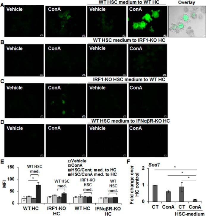
Hepatocytes from IRF1-KO and IFNαβR-KO are resistant to oxidative stress and apoptosis by ConA-stimulated WT-HSCs. A–D, hepatocytes (WT or IRF1-KO or IFNαβR-KO) were incubated in medium without or with 50 μg/ml of ConA or medium conditioned by HSCs (WT or IRF1-KO) for 8 h in the absence or presence of ConA. After 12 h of incubation, DCFDA staining was performed to determine oxidative stress in hepatocytes. An overlay of phase-contrast and DCF image in A shows ROS generation and bleb formation in WT hepatocytes treated with ConA/HSC medium from WT mice. Scale bars, 20 μm. Bar graph (E) shows mean fluorescence intensity (MFI) of combined values from three independent experiments. All images are representative of three separate experiments (see Fig. S7 for phase-contrast images). F, mRNA expression of SOD1 in WT hepatocytes incubated in medium without or with 50 μg/ml of ConA or medium conditioned by WT-HSCs. The data are representative of three sets of experiments. *, p < 0.05.
