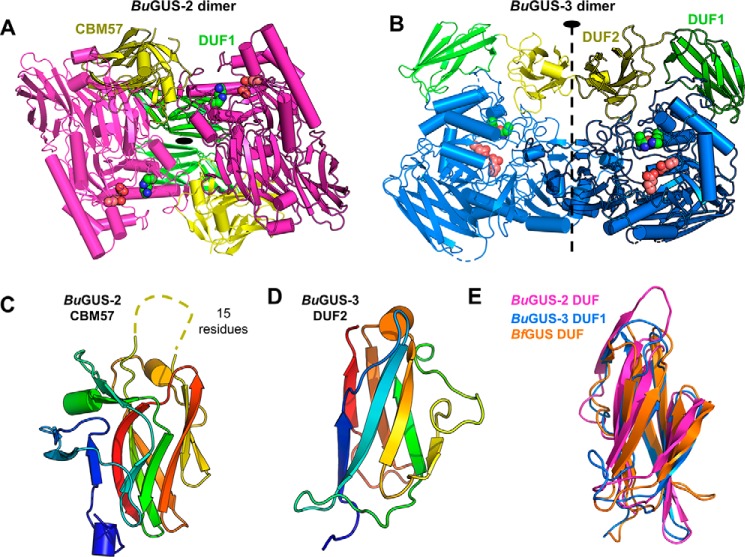
Figure 4.
Quaternary structure of BuGUS-2 and BuGUS-3 and structural analysis of C-terminal domains. A, BuGUS-2 dimer with core fold shown in magenta, DUF1 in green, and CBM57 domains in yellow with active site glutamates and the NXK motif shown as deep salmon and green spheres, respectively. B, BuGUS-3 dimer with core fold in blue, DUF1 in green, and DUF2 in yellow with catalytic glutamates and NxK motif in deep salmon and green spheres, respectively. C, CBM57 of BuGUS-2 shown with disordered loop shown as dotted line. D, structure of BuGUS-3 DUF2. E, structural alignment of BuGUS-2 DUF, BuGUS-2 DUF1, and BfGUS DUF.

