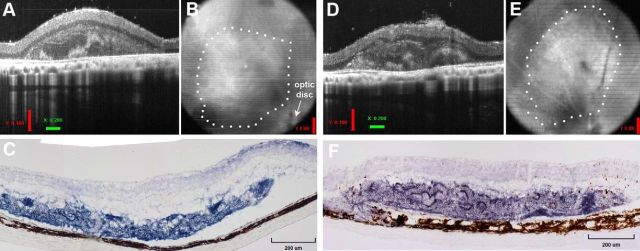
Figure 2.
Retinal transplant can survive and integrate with degenerated host retina. Examples of retinal transplants verified in vivo by high-resolution OCT at 1 month after surgery are shown for Rats R15–13 (A, B) and R15–15 (D, E). A, D, B scans show the transplant placement in the subretinal space. Regions with partial lamination are clear in addition to photoreceptor rosettes. B, E, Fundus images represent the transplant placement nasal-dorsal to the optic disc. C, F, Examples are shown of BCIP staining for human placental alkaline phosphatase (dark blue to purple) labeling donor tissue in the subretinal space at 4.4 months (C) and 3.5 months (F) after surgery in Rats R15–13 and R15–15, respectively. Dark blue visible beyond the transplant represents cells that migrated from fetal tissue and continued to develop within the host retina.

