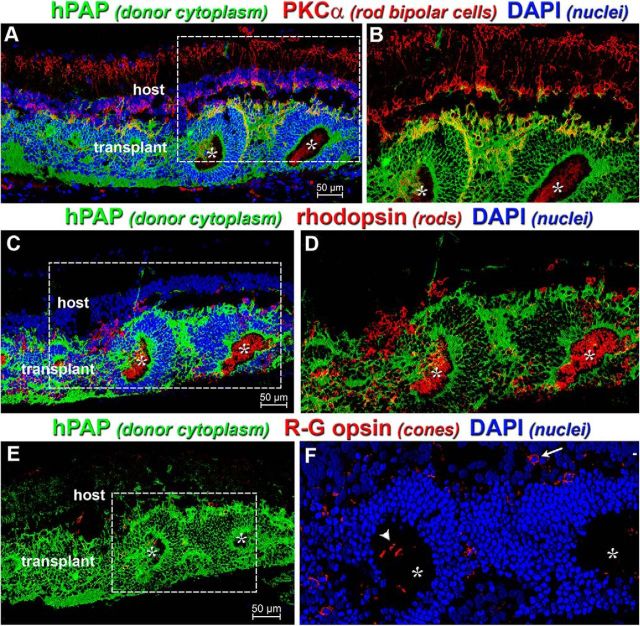
Figure 3.
Retinal transplants generate new photoreceptors and rod bipolar cells. All images represent transplant at 3.5 months after surgery from Rat R15–15 and are oriented with ganglion cell side up and RPE side down. A, PKCα is a marker of rod bipolar cells and labels both the host (hPAP−, red) and donor (hPAP+, green) tissue. Donor bipolar cells (yellow) surround photoreceptor rosettes (white asterisks) and interact with photoreceptor terminals to form a putative outer plexiform layer. B, Magnification of boxed region in A with blue channel removed. Donor bipolar cells (yellow) have synaptic projections that innervate the host inner plexiform layer. Without these connections, the photoreceptor light response will not be detected in the brain. C, Rhodopsin (Rho) expression within donor-derived photoreceptor rosettes. Rho+ outer segments (red) are indicative of functional rods and critical for light response. D, Magnification of boxed region in C with blue channel removed. Rho is localized within rosettes and the photoreceptor cell bodies (green) surround the inward-pointing outer segments. E, Red-green (R/G) opsin (red) labels cone outer segments and is mostly found within donor rosettes. There are almost no host cones remaining. F, Magnification of boxed region in E with green channel removed to allow R/G opsin signal to be more clearly seen. Cones are generated at a lower frequency than rods. Only transplant cones have outer segments (arrowhead). Arrow indicates remaining host cone.

