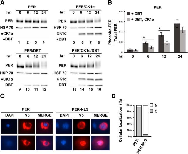
Figure 5.
CK1α antagonizes DBT activity on cytoplasmic PER. A, Drosophila S2 cells were transfected with pAc-per-V5-His together with one of the following: no kinase plasmids; pMT-ck1α-FH only, FH denotes 3XFLAG-6XHis; pMT-dbt-V5–6XHis only; or pMT-ck1α-FH and pMT-dbt-V5–6XHis. Cells were harvested and proteins extracted at the indicated times (in hours) post kinase induction. PER, DBT, and CK1α proteins were visualized by Western blotting in the presence of α-V5 or α-FLAG antibodies. Detection of α-HSP70 represents loading control. Circular dots indicate blots without any signal detected as expected. B, Quantification of phosphorylated PER (phospho-PER) using ImageLab (Bio-Rad). Following quantification, the number of phospho-PER isoforms for each time point was presented as a fraction of total PER isoforms present. Error bars indicate SEM from four biological replicates. *p < 0.05. C, Immunofluorescence of PER in S2 cells. Left, Nuclear staining using DAPI. Middle, PER staining using α-V5; (right) merged image showing localization of PER or PER-NLS. Top and bottom, Representative images of different S2 cells. D, The subcellular localization of PER and PER-NLS were determined and expressed as the percentage of total number of cells analyzed per construct (n = 34). N, Nuclear; C, cytoplasmic localization.

