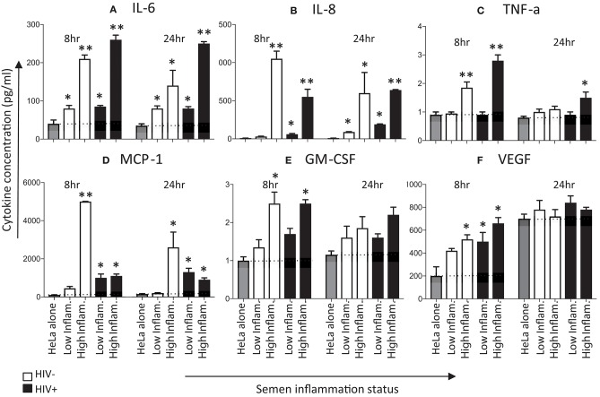
Figure 3.
Effects of seminal plasma on cytokine secretion by HeLa cells. Hela cells were exposed to seminal plasma from HIV-negative (white bars) or HIV-infected individuals (black bars). A 1:50 dilution of seminal plasma from men with extreme inflammatory profiles (highest vs. lowest percentile) were used to stimulated HeLa cells for 8 and 24 h. Cytokine levels was measured in the 8 and 24 h post-stimulation in Hela cells supernatants by Luminex. Seminal plasma (1:50) in serum free culture media was used, while control was serum free culture media. Data are represented as the mean ± SEM of duplicate wells in each experiment. Wilcoxon matched pairs (non-parametric) and a standard matched pairs t-tests (parametric) were used to compare the control and treatment groups. *p < 0.05; **p < 0.01. (A) IL-6, (B) IL-8, (C) TNF-α, (D) MCP-1, (E) GM-CSF, and (F) VEGF.

