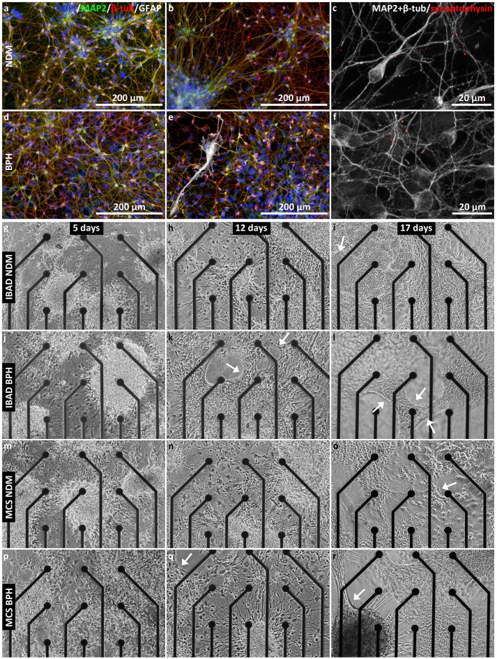
Figure 5.
Neuronal networks in NDM and BPH media. (a–f) Immunocytochemistry images from neuronal networks in NDM (a–c) and BPH (d–f) after 12 days in adherent culture. Networks in both media contained MAP2 and β-tub-positive neurons, whereas GFAP-positive astrocytes were only found in BPH medium (e). Synaptophysin was expressed in networks in both media (c,f). (g–r) Development of the network in NDM (g–j,m–o) and BPH media (j–l,p–r) on BMT MEAs (g–l) and MCS MEAs (m–r) at 5, 12, and 17 days in adherent culture. Network retraction (indicated by arrows) started earlier in BPH (k and q) than in NDM (i and o) on both MEA types.

