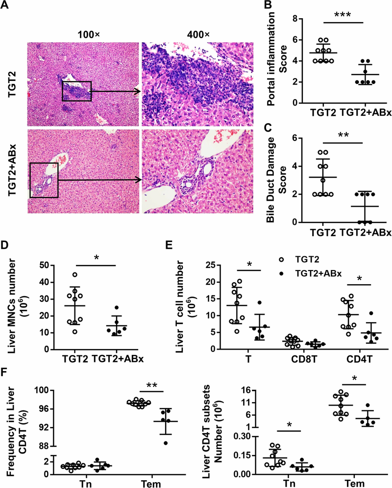
Figure 5. Antibiotic treatment alleviated exacerbated cholangitis in dnTGFβRII TLR2−/− mice.
(A) Representative H&E staining of liver sections of 13–14-week-old female TGT2 mice with (TGT2+ABx) or without (TGT2) mixed antibiotics treatment. (B) Scores of portal inflammation in TGT2 (n=9) and TGT2+ABx mice (n=7). (C) Scores of bile duct damage in TGT2 (n=9) and TGT2+ABx mice (n=7). (D) Absolute number of hepatic mononuclear cells from TGT2 (n=9) and TGT2+ABx mice (n=6). (E) Cell numbers of total T cells and CD8+T and CD4+T cell subsets in the liver of TGT2 (n=9) and TGT2+ABx (n=6) mice, as determined by flow cytometry. (F) Frequency (left) and absolute number (right) of naive CD4+T (CD44-CD62L+) and effector memory CD4+T (CD44+CD62L-) cell subsets in the liver of TGT2 (n=9) and TGT2+ABx (n=6) mice. Data are presented as mean ± SD. *p<0.05, **p<0.01, ***p<0.001.

