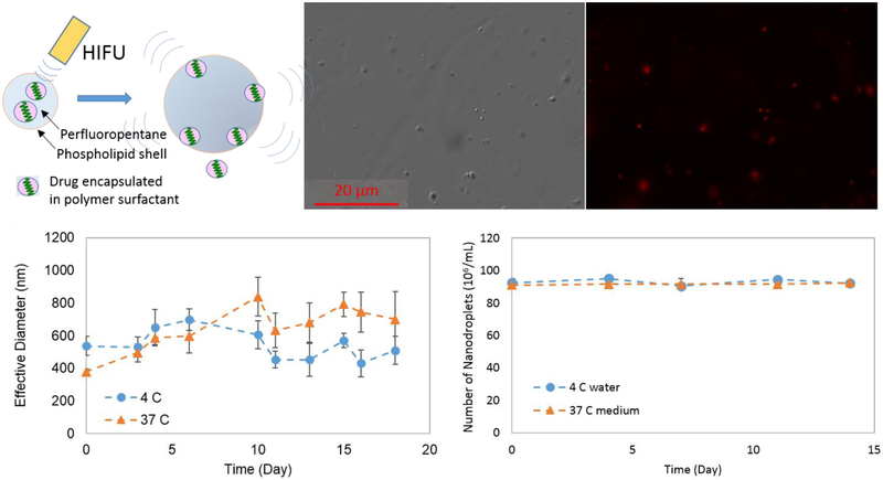
Figure 1.
(A) Schematic of drug release from the nanodroplet via phase-transition by HIFU trigger. (B) DIC (Differential Interference Contrast) and fluorescence optical images (63x magnification). (C) Effective (hydrodynamic) diameter measured by DLS over time at two different temperatures. (D) The number of nanodroplets counted over time at 4 °C and at 37 °C in the medium using phase-contrast.

