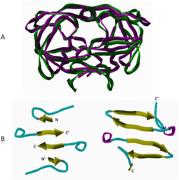
Figure 2. Comparison of the overall structures and dimer interfaces of HIV-1 and XMRV proteases.
(A) Superposition of overall structures of HIV-1 PR (magenta) and XMRV PR (green) using ribbon/tube representation. (B) Comparison of dimer interface regions of HIV-1 PR (left) and XMRV PR (right) using ribbon/tube representation (yellow: beta-sheet, cyan: loop, magenta: alpha-helix). The N- and C-terminal ends of the monomers are indicated.

