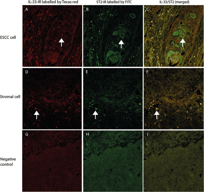
Fig. 4.
Double immunofluorescence (DIF) staining with confocal microscopy to evaluate the co-expression of IL-33 with its receptor, ST2, in the ESCC section. DIF images showed that IL-33-IR (labelled by Texas red, red cells in A, D) was frequently co-localized (merged images in C, F) with ST2-IR (labelled by FITC, green cells in B, E) in ESCC cells (merged image in C) and stromal cells (merged image in F). IRs for targeted proteins were not shown in isotopy-matched negative controls (G–I). (A–I: DIFs, original magnification ×200; counterstaining was not applied)

