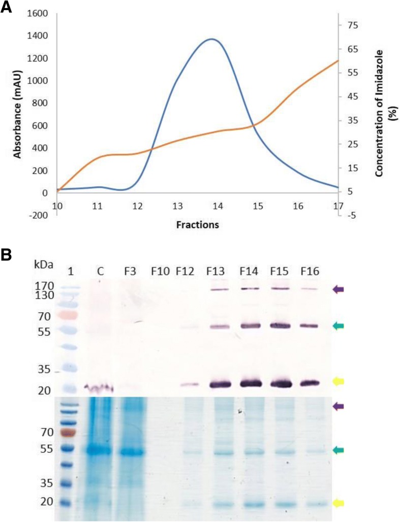
Fig. 3.

Purification of the N-protein using nickel affinity chromatography. a Chromatographic trace showing 6xHis-tag N-protein elution (orange line) from the 6xHis-tag-chelating affinity column with increasing imidazole concentration (blue line). b Western blot analysis (top) and Coomassie-stained gel (bottom) of collected fractions. Lane 1 contained PageRuler ™ Prestained protein ladder (Thermo Scientific, MA, USA), C contains crude plant extract, F3 represents unbound wash fraction 3, F10 contained a wash fraction 10, F12–16 protein peak visualised in fractions 12–16. The protein was detected with 1:5000 anti-N primary antibody and 1:5000 anti-rabbit secondary antibody. The purple arrow indicates the pentamer, the green arrow indicates the dimer and the yellow arrow indicates the monomer
