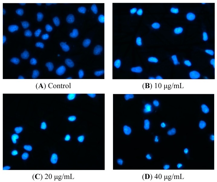
Figure 3.
Nuclear morphologic changes in apoptotic HeLa cells by tatariside G. The cells were dyed with Hoechst 33258 after 12 h of treatment with tatariside G (10, 20, and 40 μg/mL). (A) Normal nuclei are symmetrical baby blue. (B) Intact nuclei are asymmetrical bright blue in the early stage of apoptosis. (C) and (D) Fragmented nuclei are presented in the advanced stage of apoptosis. Magnification: 400×.

