Figure 4.
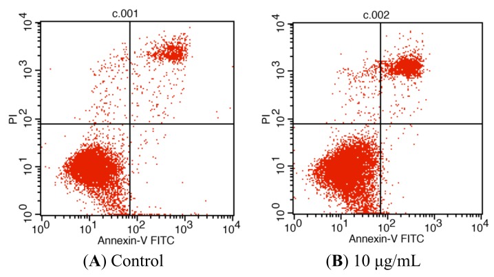
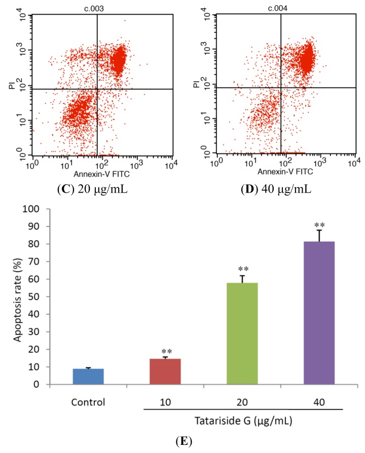
Tatariside G-induced apoptosis of HeLa cells was discriminated by AV and PI double staining. Following 24 h of treatment with tatariside G, the cells were labeled with AV and PI and analyzed by flow cytometry. The lower left quadrant shows vital cells (double negative) and the lower right quadrant indicates early apoptotic cells (AV positive but PI negative). The upper right quadrant represents late apoptotic cells (double positive). (A)–(D) Typical images. (E) Average apoptosis rate of cells in each group. ** p < 0.01 vs. the control group. Data are expressed as the mean ± SD. n = 3.


