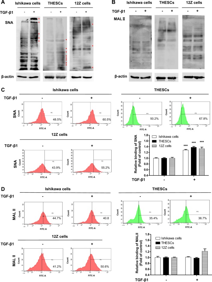
Fig. 2. α2-6 Sialylation of endometrial cells is increased by TGF-β1 treatment.
Ishikawa, THESCs, or 12Z cells were incubated with TGF-β1 for 48 h. The expression of α2-3 or α2-6 sialic acid epitopes in endometrial or endometriotic cells was determined by lectin blot a, b or lectin FACS analysis c, d using biotin-labeled MAL II and SNA, respectively. Data from the lectin FACS analysis are expressed as the mean ± SD of three independent experiments. Red asterisk indicates increased protein expression in the TGF-β1-treated group compared with the control group. * p < 0.05 and *** p < 0.001 compared with the control group

