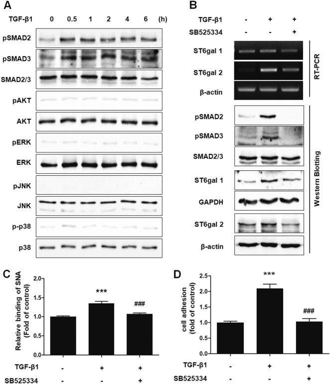
Fig. 4. TGF-β1 induces the adhesion of endometrial cells to mesothelial cells through the TGF-βRI/SMAD2 signaling pathway.
a Ishikawa cells were cultured with TGF-β1 (10 ng/mL) according to the indicated times. Phosphorylation levels of SMAD2, SMAD3, Akt, ERK, JNK, and p38 were examined by western blot analysis. b–d Ishikawa cells were treated with 10 μm TGF-βRI inhibitor (SB525334) 1 h before TGF-β1 (10 ng/mL) stimulation. b Phosphorylation of SMAD2 and SMAD3 was determined 4 h after TGF-β1 treatment. Expression levels of ST6Gal1 and ST6Gal2 were estimated 24 h after TGF-β1 stimulation. c Ishikawa cells were incubated with TGF-β1 for 48 h. Using biotin-labeled SNA, α2-6 sialic acid epitopes in Ishikawa cells were analyzed via FACS using biotin-labeled SNA. d Tracker™ Green CMFDA-labeled Ishikawa cells were added onto Met-5A cells 48 h after TGF-β1 treatment. After incubation and washing, the number of attached Ishikawa cells was calculated and is expressed as the fold-difference relative to the control (mean ± SD). ** p < 0.01 for comparison between two groups. ##p < 0.01 compared with the TGF-β1-treated group

