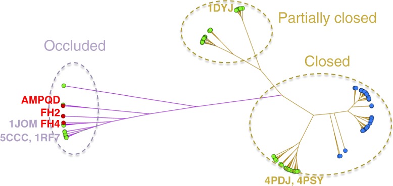
Fig. 8.

Clustering of 162 DHFR structures based on their pairwise Cα RMSD of the Met20 loops. DHFR structures are represented by circles filled in blue (humans), green (eDHFR), and red (in this study). The edge length (colored in purple for the occluded and gold for the closed conformations, respectively) is proportional to the maximum RMSD of the Met20 loop conformers. Please see a more detailed clustering diagram in the Supplementary Information
