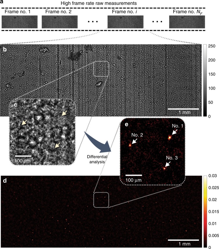
Fig. 4. Experimental demonstration of applying the CMA algorithm and OFN to a lysed blood sample spiked with motile trypanosome parasites, over an FOV of ~14.7 mm2.
a A time-sequence of the diffraction patterns of the sample is captured. b, c One of the frames in the raw image sequence is shown. The diffraction pattern is severely speckled due to the light scattering by the cell debris, which renders the parasites invisible (yellow arrows in (c)). d, e After being processed by the CMA algorithm, motile parasites can be detected. The amplitude and phase movies of the three trypanosomes in e (indicated by white arrows) are also shown in Supplementary Movie 2

