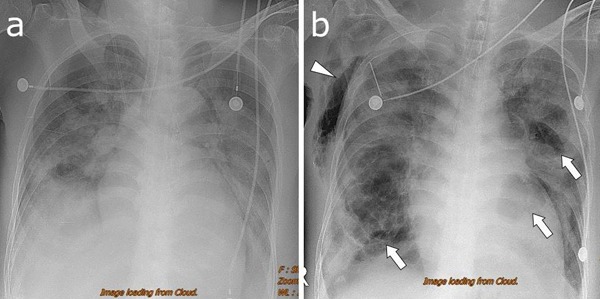
Figure 1.

(A) Chest radiography on hospital day 7 reveals a diffuse consolidation of the left lung and patchy areas of the consolidation in the right lung. (B) Radiography on hospital day 12 shows multiple cavities of variable sizes within the consolidation of the bilateral lungs (arrows). Subcutaneous emphysema of the right chest can be observed (arrowhead). A needle is inserted in the right upper intercostal space.
