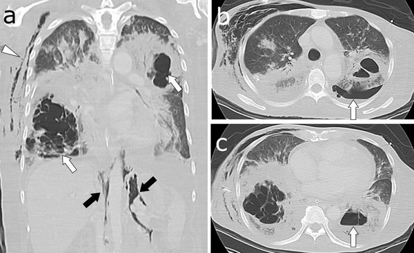
Figure 2.

(A) Lung window of a CT shows necrotizing pneumonia in the right lower and left upper lobes (white arrows). Subcutaneous emphysema (arrowhead) and pneumoperitoneum (black arrows) can be observed. (B) An air-fluid level is present in the left pleural space (arrow), representing pyopneumothorax. (C) A cavity contains an air-fluid level within the left lower lobe consolidation (arrow), suggesting lung abscess. CT, computed tomography.
