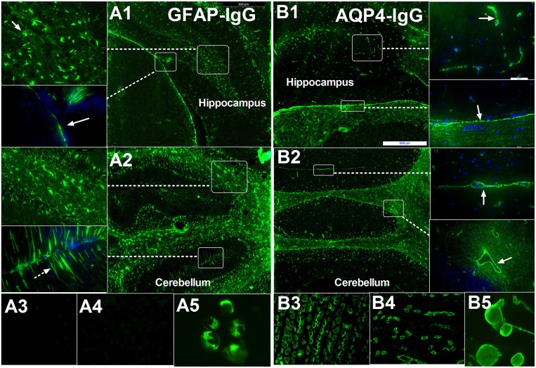
Figure 1.
Comparison of immunofluorescence-pattern between GFAP-IgG and AQP4-IgG. (A1–A5) IgG from patient with GFAP astrocytopathy. (A1) The IgG is bound to the foot process at pia (arrow) and astrocyte body in hippocampus. (A2) IF pattern in different layer of cerebellum: (1) The IgG is bound to the astrocyte body in different layer, especially white matter (arrow); (2) it was detected in molecular layers with bergmann radial pattern (arrow). (A3,A4) no pattern-specific staining was detected in the kidney and stomach tissue but positive for GFAP-transinfected HEK-293 cell. (B1–B5) IgG from a positive AQP4-IgG NMO patient. (B1) The IgG is bound to the cell with the foot process around the microvessels (arrow) in brain and pia (arrow). (B2) Anti-AQP4 pattern located at the border between two molecular layers with Virchow-Robin space profile (arrow) without bergmann radial pattern. (B3–B5) Pattern-specific staining was detected in the stomach, kidney tissue, and AQP4-transinfected HEK-293 cell.

