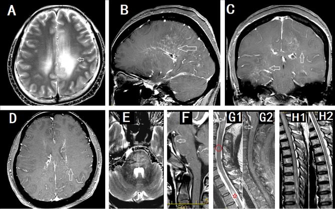
Figure 2.
Imaging findings in patients with GFAP astrocytopathy. (A–D) were from a female meningoencephalitis patient. (A) MR images showing extensive abnormalities in the white matter around the ventricle (arrow). (B) Sagittal section showed linear perivascular radial gadolinium enhancement in the white matter perpendicular to the ventricle(arrow). (C,D) Coronal section (C) and cross section (D) showed vessel-like enhancement (arrows). (E) and (F) from a male meningoencephalitis patient showed pons abnormality (black arrow) and pia enhancement (white arrow). (G1) and (G2) were from a female with myelitis. (G1) Cervical lesion extended to the area postrema of medulla(arrow), sparse cervical abnormality(red round area) and thoracic LESCLs (star marker). (G2) Slightly enhancement in medulla (arrow). (H1) and (H2) were from a male meningoencephalomyelitis patient. Longitudinal extensive lesions in the whole spinal cord (H1) and soon recovery after the treatment (H2).

