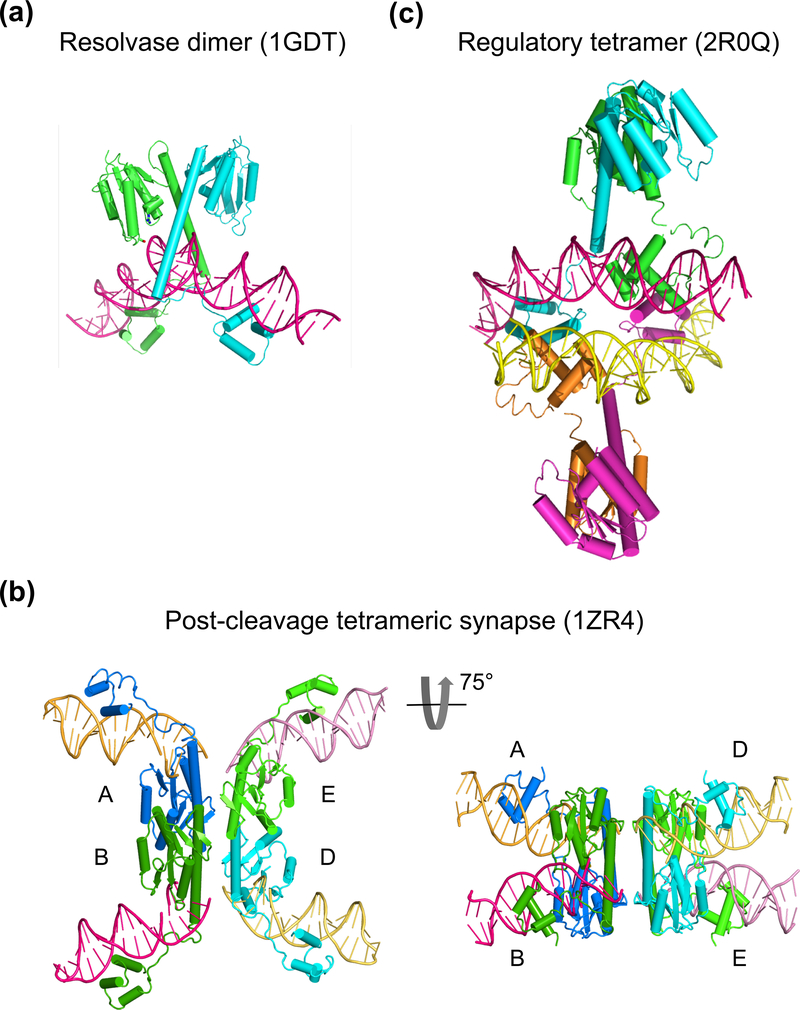
Fig. 6.
Crystal structures of resolvase-DNA complexes. (a) The pre-cleavage resolvase dimer-site I DNA complex (PDB: 1GDT). The protein subunits are shown as cyan and green ribbon diagrams and DNA in pink tube-and-ladder. (b) Two views of a post-cleavage tetrameric resolvase complex with two cleaved DNAs (PDB: 1ZR4). On the left is the conventional view with DNAs appearing to be outside of the protein tetramer. On the right is a view rotated about the horizontal axis by 75°. The diagonal subunits in the left panel are now side-by-side, and the light and dark pink as well as yellow and orange DNAs appear to be co-linear. (c) A synapse of Sin resolvase tetramer bound to two regulatory site DNAs (PDB: 2R0Q). DNAs (pink and yellow) are inside of the protein tetramer (cyan, green, orange and magenta).

