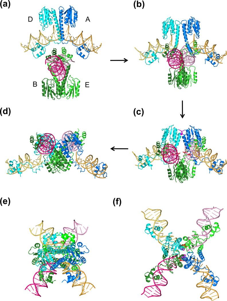
Fig. 8.
Strand passage mechanism. (a) A diagram of topoisomerization by a type II enzyme. The dimeric Topo II protein itself has two gates (N and C), which can open and close in an ATP hydrolysis and DNA dependent manner. DNA T segment (shown in grey) is transported through the open protein N gate, the cleaved DNA gate (G segment, shown in green, and eventually out of the protein C gate. This figure is a reproduction of Schoeffler et al. (Schoeffler and Berger, 2008) with permission. (b) A diagram of SRs recombining DNAs by the strand passage mechanism. Each SR subunit contains a large N-terminal catalytic domain and a small C-terminal DNA binding domain linked by the long E helix. Two SR dimers (in distinct colors) enclose the two DNA recombination sites (grey and green). Polarity of res DNA is indicated by the arrowhead. DNA cleavage requires some conformational changes at the dimer interface. After DNA cleavage, the two SR dimer-cleaved DNA complexes approach each other and DNAs cross and pass each other through the cleaved DNA and the open C-termini of E helices. The two DNAs are recombined from being horizontally connected at the beginning to vertically connected, which is reminiscent of recombination by YRs as diagramed in Fig. 2b. (c) A topological diagram of DNA resolution by Tn21-family resolvases. The two res sites (direct repeats) are shown in green and blue and the intervening DNAs shown in light brown. Each res site consists of site I, II and III as labeled. The recombination synapse traps 3 negative supercoils as experimentally determined. Additional local + and - supercoil occur but cancel one another. After strand cleavage, crossing and partner switching, the ligation product two singly linked catenanes.

