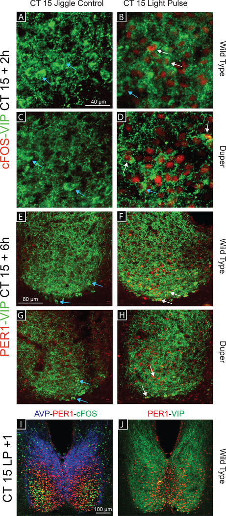
Figure 3.
Photomicrographs illustrating immunostaining for cFOS-VIP, PER1-VIP, and AVP-cFOS-PER1. (A-D) Representative images of a double labeled SCN in Experiment 1. cFOS-ir (red) and VIP-ir (green) are shown in WT (A, B) and duper (C, D) hamsters 2h after either a cage movement (control procedure, A, C) or a 15-min light pulse (B, D) at CT15 (40X). (E-J) Double labeled SCN in Experiment 2. PER1-ir (red) and VIP-ir (green) are detected in WT (E, F) and duper (G, H) hamsters 6h after either a cage movement (E, G) or a 15-min light pulse (F, H) at CT15 (20X). White arrows indicate VIP co-labeled cells; light blue arrows indicate cells stained for VIP only. (I, J) Representative 10X images of WT SCN 1h after a light pulse at CT15 triple labeled (I) for AVP (blue), cFOS (green) and VIP (red) or double labeled (J) for PER1 and VIP. Note that PER1-ir cells are medial to cFOS-ir cells.

