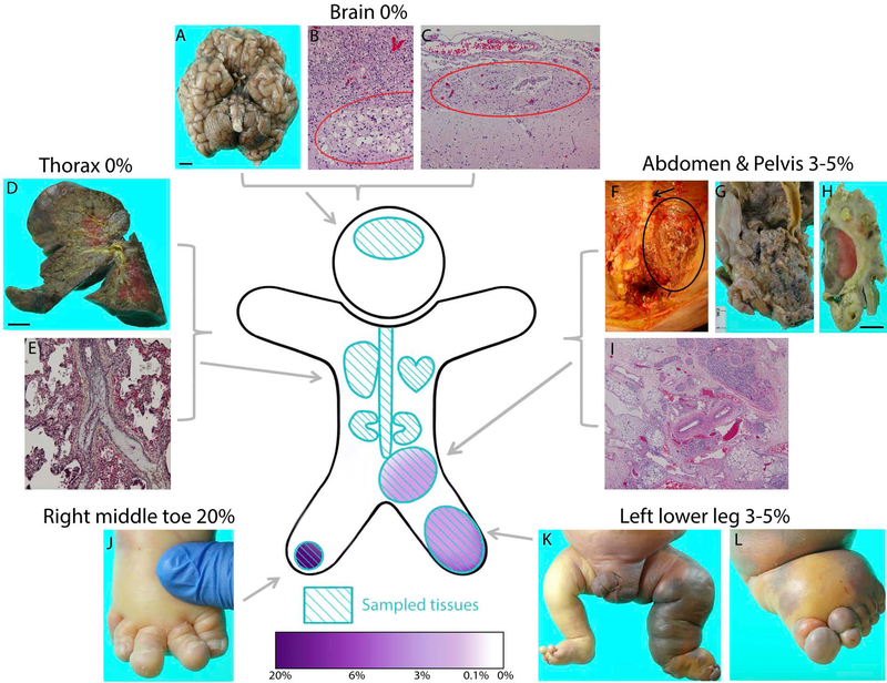
Figure 1. PIK3CA p.E545K mutation levels and autopsy findings:
Black scale bars correspond to 1 cm. Tissues sampled for ddPCR testing (light blue) and presence of PIK3CA muation (purple) represented in cartoon figure. Not all sampled tissues are pictured. A: external gross brain anatomy indicates normal development except for generalized cerebellar atrophy. Brain size smaller than average (390 gm). B: Periventricular leukomalacia (circle). C: Leptomeningeal glioneuronal heterotopia (circle) near R thalamus. D: Right lung demonstrating chronic neonatal lung disease and E: remote thromboembolus (central vessel with luminal fibrous occlusion). F: Pelvic and retroperitoneal extension of lymphatic malformation (circle) near spine (arrow) G: Posterior view of vascular malformation near bifurcation of abdominal aorta, encasing left common iliac. H: Vascular malformation surrounding left kidney. I: The rectum is surrounded by loose fibrous tissue containing variably-sized vascular spaces and small islands of adipose and lymphoid tissue with hemorrhage/congestion. J: Right foot exhibiting macrodactyly of toes two, three, and four. K: Significant overgrowth and dusky, violaceous discoloration seen in left leg. L: Left foot exhibiting overgrowth, edema, and discoloration.

