Figure 1.
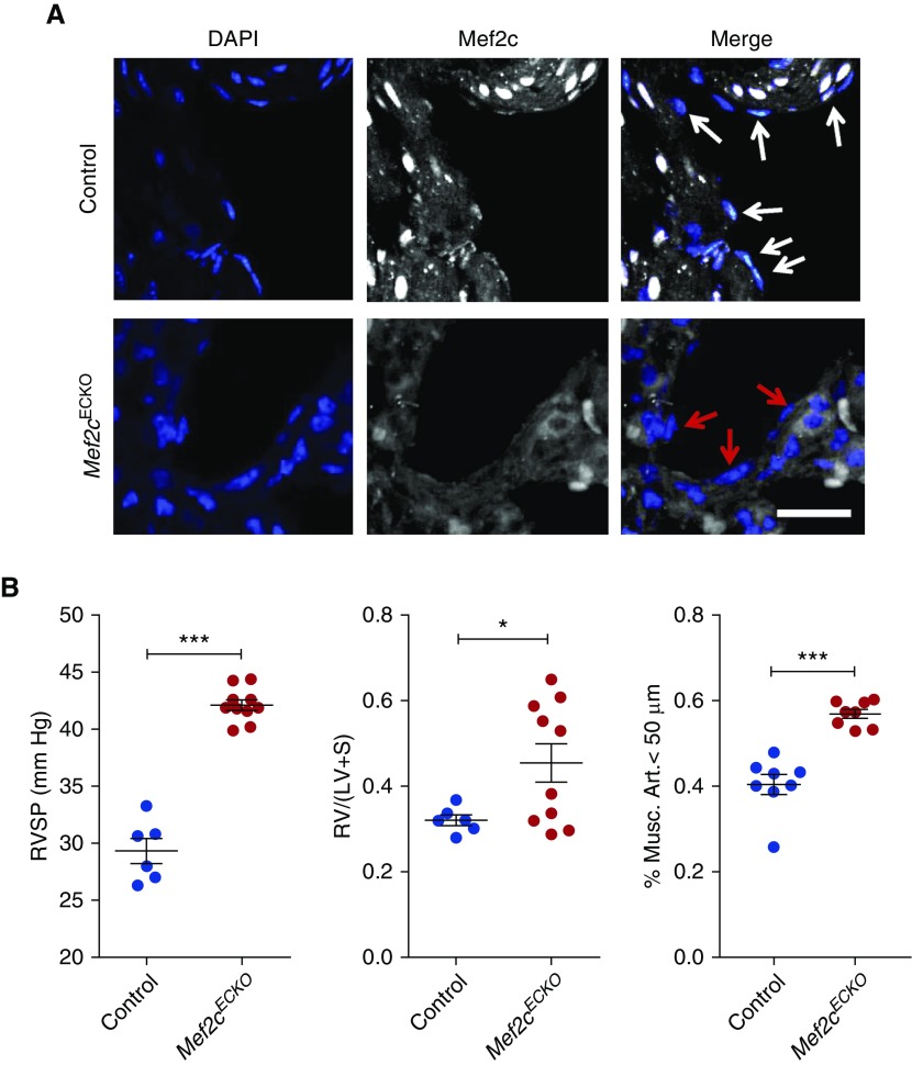
Endothelial-specific deletion of Mef2c leads to worsening pulmonary hypertension. (A) Immunofluorescent staining for Mef2c (white) in the lungs of Mef2cECKO mice. White and red arrows designate endothelial cell nuclei. DAPI (blue) is also shown. Scale bar: 50 μm. (B) (Left) Right ventricular systolic pressure (RVSP) measurements of control and Mef2cECKO mice after exposure to chronic hypoxia. ***P < 0.001. n = 6–10 per group. (Middle) Right ventricle to left ventricle + septum [RV/(LV + S)] weight ratios for control and Mef2cECKO mice after exposure to chronic hypoxia. *P < 0.05. (Right) Quantification of muscularized pulmonary vessels in Mef2cECKO mice and controls. ***P < 0.001. Mef2c = myocyte enhancer factor 2c; Musc. Art. = muscularized pulmonary arterioles.

