
Figure 5.
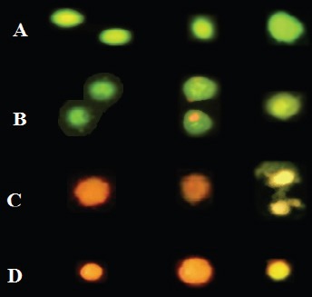
Multiple Images of Cell Death Stained with AO/PI to Compare Their Features. A) Normal viable cells are bright green. Various pictures of apoptotic cells selected from cultures are given in B & C. B) The viable apoptotic cells in earliest stage possess a fragmented and condensed nucleus with circumnuclear chromatin condensation that emerges as a bright green area and even cells may have apoptotic bodies around cytoplasm as are shown. Yellow/orange and condensed nuclei are apoptotic cells which enter the late stage. C) Dead cells with condensed and shrieked nuclei are late apoptotic orange cells. D) Necrotic cells have orange/red nuclei with an organized structure.