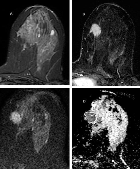
Figure 1.

A 35 Years Old Women with Palpable Left Breast Lower Inner Quadrant Spiculated Mass. The mass has low T2 signal (A), homogenous enhancement with spiculated border in post contrast image (B) and is restricted in DWI with ADC value 1.02 × 10⁻³ mm2/s (C). Two methods of ROI placement is depicted in ADC image (D). The pathology was invasive lobular carcinoma.
