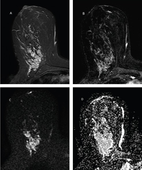
Figure 2.

A 35 Years Old Women with Palpable Large Right Breast Lower Inner Quadrant Non-mass Lesion. The lesion has high T2signal (A), clumped pattern enhancement in post contrast image (B) and is mildly restricted in DWI with ADC value 1.60 × 10⁻³ mm2/s (C). Two methods of ROI placement is depicted in ADC image (D). The pathology was ductal carcinoma in situ.
