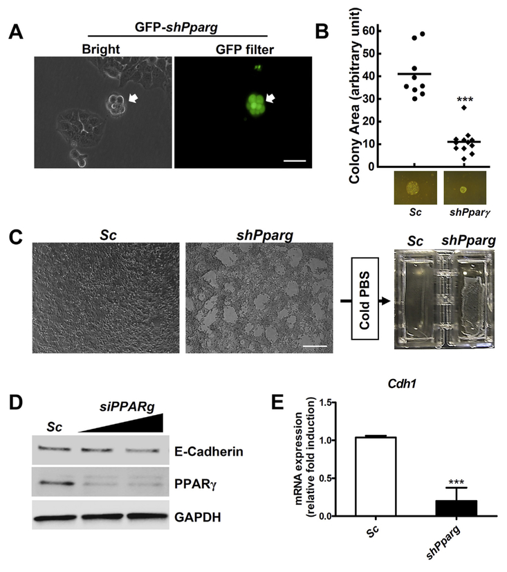
Fig. 4.
Cellular adhesion of MLE-15 cells was reduced by knock-down of Pparg gene. (A) Colony shape of MLE15 cells is shown after transfection with shPparg plasmid for 4 days. Transfected cells were visualized by turbo GFP (tGFP) protein. Scale bars: 100 μm. (B) Colony areas from stable MLE-15-shPparg and parental cells. (C) MLE-15-shPparg and parental cells were grown to 100% confluence and challenged with cold PBS. Lifted cells are shown in MLE-15-shPparg cells. Scale bars: 300 μm. (D) MLE-15 cells were transfected with siPparg or scramble RNA and total protein extracts were subjected to Western blotting. (E) Expression of Cdh1 in MLE-15-shPparg cells was measured by qRT-pCR. Gapdh mRNA was used as internal control. ***, p < 0.0001.

