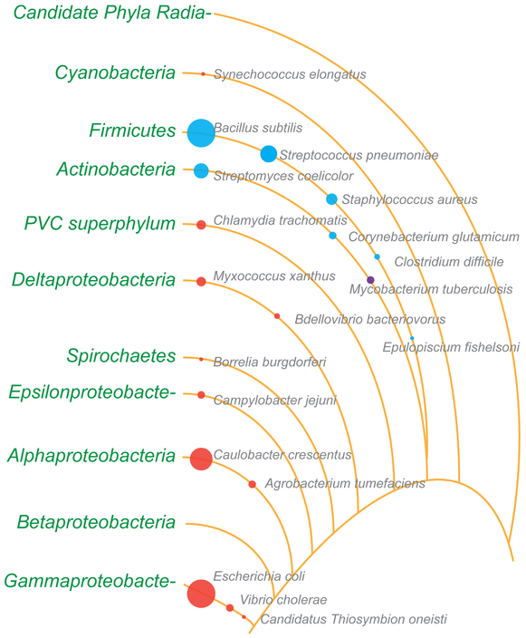
Figure 1.
Representation of the relative number of reports describing cell division in various bacterial species. The diameters of the circles roughly indicate the number of cell division publications available for organisms highlighted in this review. Note: The diameter of the circles for E. coli and B. subtilis are capped at an arbitrary number so that other circles are visible. Red circles, Gram-negative; blue circles, Gram-positive; violet, M. tuberculosis. Lines depict phyla and lineages loosely based on the bacterial branch of the tree of life (PMID: 27572647). Several phyla and lineages were omitted for clarity. PVC superphylum comprises of Planctomycetes, Verrucomicrobia, and Chlamydiae. The branch length, spaces between them, and the order in which some organisms are listed are not based on phylogeny.

