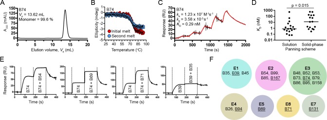
Fig 2. Biophysical characterization of VHHs.
Representative SEC profile (A) and thermal unfolding curve from initial and refolded thermal melts (B). (C) Representative SPR single-cycle kinetics sensorgram showed high-affinity binding of VHHs to biotinylated TcdB1751-2366 immobilized on a CAP sensor chip. (D) Plot comparing KDs of VHHs isolated from solution and solid-phase panning schemes. The four VHHs isolated from both panning schemes were included in the analyses. Bars represent the median KD and a P-value < 0.05 was considered significant (Mann Whitney two-tailed unpaired t-test). (E) Representative sensorgrams demonstrating SPR-based epitope binning. All VHHs were injected at 10x KD concentrations. (F) Summary of the TcdB1751-2366 epitope bins identified in this study by the pool of VHHs tested. The VHHs in each epitope (E) bin are noted and the underlined VHHs represent the highest affinity and/or slowest dissociating antibodies in each bin.

