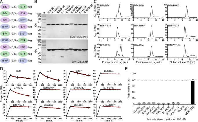
Fig 3. Generation and characterization of VHH-VHH dimers.
(A) Cartoon diagram of VHH-VHH dimers separated by a 25 amino acid linker (G4S)5 and containing a C-terminal His6 “tag”. (B) Dimers were expressed, purified and analyzed by non-reducing (NR) SDS-PAGE and probed by α-His6-AP in Western blot (WB). M, protein molecular mass marker. (C) SEC profiles of dimers. Asterisks denote the peaks that were selected for SPR analysis when aggregates were present. (D) SPR sensorgrams showing dissociation phases of VHH-VHHs and parent monomers. (E) TcdB neutralization with VHH-VHH dimers. The final TcdB concentration of 3–10 pM and VHH-VHH concentration of 1 μM was co-incubated with Vero cell monolayers for 72 h before addition of WST-1 cytotoxicity reagent. MDX-1388 was used at 250 nM. Neutralization values are presented as mean ± SD from n = 3 independent experiments.

