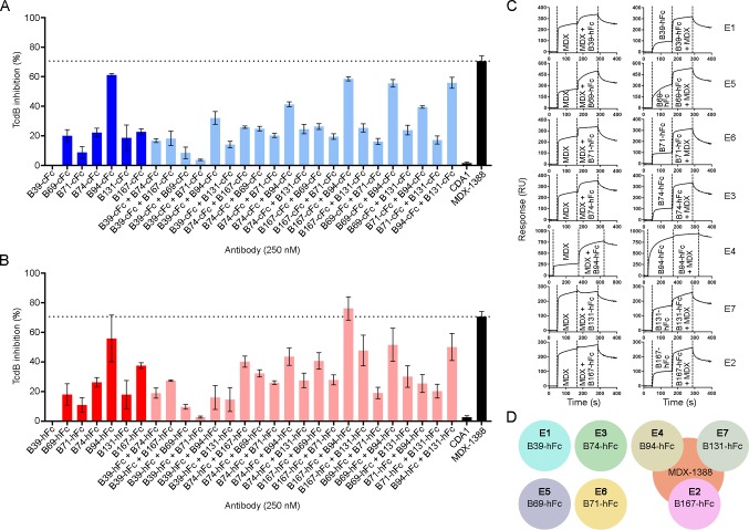
Fig 5. TcdB neutralization assays with VHH-Fcs.
(A) TcdB neutralization with VHH-Fcs containing a camelid hinge. (B) TcdB neutralization with VHH-Fcs containing a human hinge. In (A) and (B), the final TcdB concentration of 500 fM and final antibody concentration of 250 nM (single antibody) or 125 + 125 nM (pairs) were co-incubated with Vero cell monolayers for 72 h before addition of WST-1 cytotoxicity reagent. Neutralization values are presented as mean ± SD from n = 4 independent experiments. (C) Sensorgrams showing SPR-based epitope binning of MDX-1388 mAb with each VHH-hFc, in both orientations, with antibodies injected at 25–50 × KD concentrations over TcdB1751-2366 surfaces. Epitope bins corresponding to each VHH-hFc are noted. (D) Summary of VHH-Fc reactivity to TcdB1751-2366 illustrating that several VHH-Fcs bind TcdB at sites distinct from MDX-1388, while others bind TcdB at regions that partially overlap with MDX-1388.

