Figure 1.

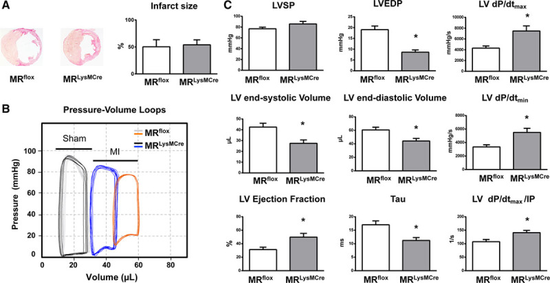
Mice with myeloid cell–restricted MR (mineralocorticoid receptor) deficiency display improved cardiac function and remodeling after myocardial infarction (MI). A, Representative sections from MRflox and MRLysMCre infarcted hearts and infarct size. B, Representative left ventricular (LV) pressure-volume loops measured in vivo with conductance catheter in sham-operated MRflox (gray) and MRLysMCre (black) mice and in MRflox (orange) and MRLysMCre (blue) mice with MI. C, LV systolic pressure (LVSP), LV end-diastolic pressure (LVEDP), LV end-systolic and end-diastolic volumes; LV ejection fraction, LV maximal rate of pressure rise (LV dP/dtmax), maximal rate of pressure decline (LV dP/dtmin), and LV dP/dtmax divided by instantaneous pressure (IP) and the time constant of LV pressure isovolumic decay (Tau). Mean±SEM (n=14–16). *P<0.01 vs MRflox.
