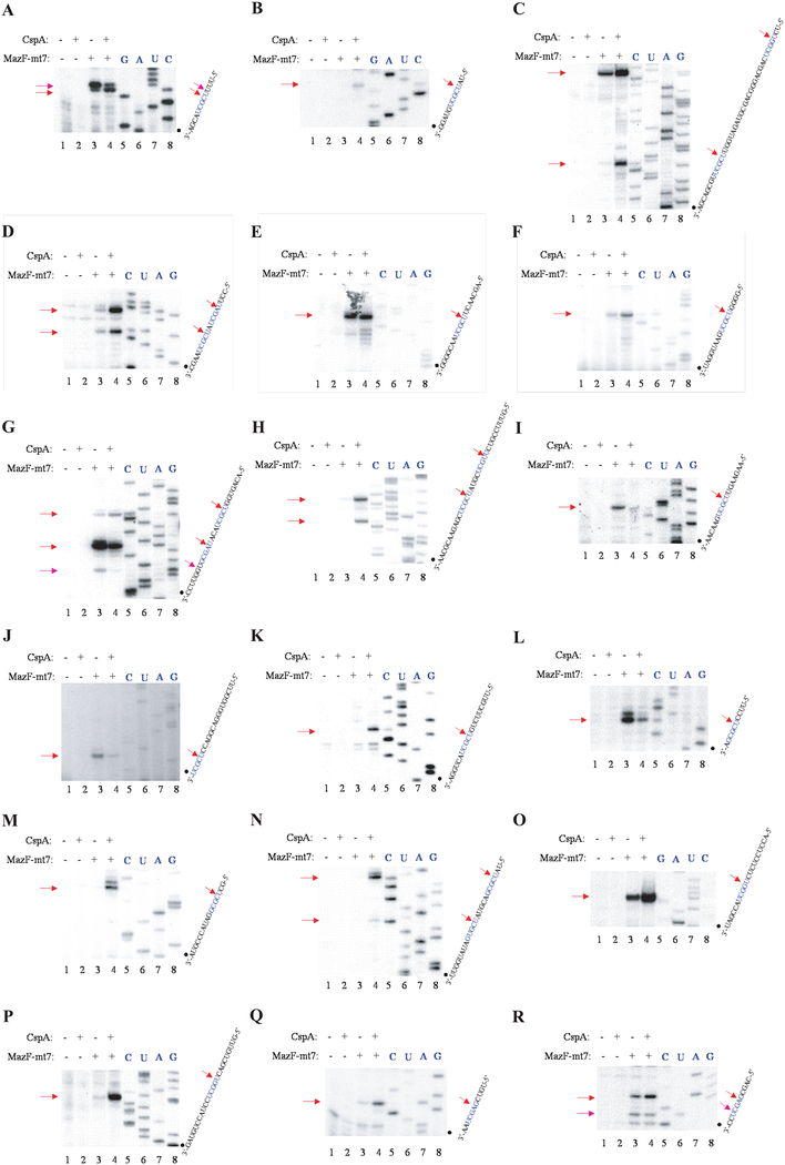
Figure 4. Cleavage of MS2 RNA by MazF-mt7.
A-O, In vitro cleavage of the MS2 RNA with (His)6MazF-mt7. Lane 1, control reaction in which no enzymes were added; lane 2, control reaction in which only CspA protein was added; lane 3, MS2 RNA incubated only with (His)6MazF-mt3; lane 4, MS2 RNA incubated with (His)6MazF-mt3 and CspA protein. Cleavage sites are indicated by red arrows (strong cleavage sites) or pink arrows (weak cleavage sites) on the RNA sequence and were determined using the RNA ladder shown on the right.

