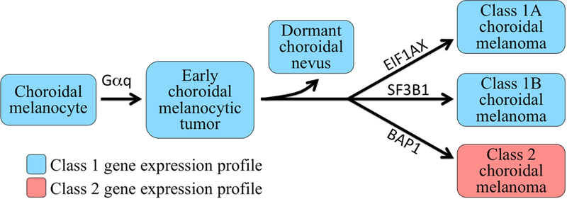
FIGURE 3.

Schematic diagram depicting current understanding of how choroidal melanoma progresses. First, choroidal melanocytes acquire a Gαq mutation that leads to development of an early choroidal melanocytic neoplasm.58,59 The vast majority of such nevi are arrested by tumor suppressor and/or immune surveillance mechanisms and driven into a dormant state. Some lesions progress past this checkpoint to the “BSE node,” in which they acquire mutations in either BAP1 (BRCA1 Associated Protein 1), SF3B1 (Splicing Factor 3B Subunit 1), or EIF1AX (Eukaryotic Translation Initiation Factor 1A, X-Linked), in a mutually exclusive manner. Tumors that acquire an SF3B1 or EIF1AX mutation retain the class 1 profile (class 1A or class 1B, respectively), whereas those that undergo BAP1 inactivation acquire a class 2 profile. Gαq mutations include mutual exclusive hemizygous activating point mutations in GNAQ (G protein subunit alpha Q), GNA11 (G Protein Subunit Alpha 11), PLCB4 (Phospholipase C Beta 4) and CYSLTR2 (Cysteinyl Leukotriene Receptor 2).
