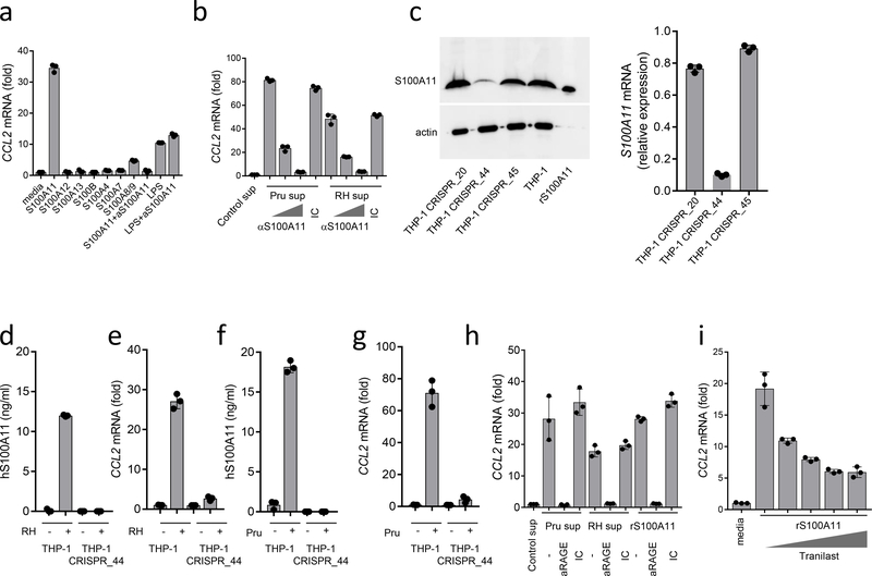
Figure 4.
Role of RAGE in S100A11-induced CCL2
THP-1 cells were stimulated with purified recombinant S100A11, S100A12, S100A13, S100B, S100A4, S100A7, S100A8/9 (all at 10 ng/ml) or with LPS (1 μg/ml), and CCL2 expression was analyzed by RT-PCR 12 hours after stimulation. In several experiments, an anti-S100A11 antibody was added to S100A11 protein (S100A11+α−S100A11) or to LPS (LPS+α−S100A11) for 30 min prior to stimulation. (b) Cell culture supernatants were collected from uninfected (control), Pru- or RH88-infected human fibroblasts and were used to stimulate monocytes directly or in the presence of increasing amounts of anti-S100A11 antibody or the isotype control (IC). CCL2 expression was analyzed by RT-PCR. (c) The knockdown of S100A11 in THP-1 cells (THP-1_CRISPR_44) was revealed by immunoblot and qRT-PCR and resulted in loss of S100A11 release and induction of CCL2 in response to (d,e) RH88 and (f,g) Pru infections. (h) Induction of CCL2 by S100A11 or cell culture supernatants collected from T. gondii-infected cells was prevented by adding an anti-RAGE antibody but not the isotype control (IC), (i) Induction of CCL2 by S100A11 was partially blocked by tranilast. The data shown represent the mean ± SD of assays performed in triplicate and are representative of four independent experiments performed.

