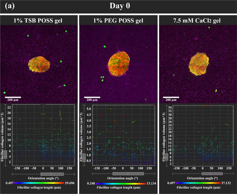
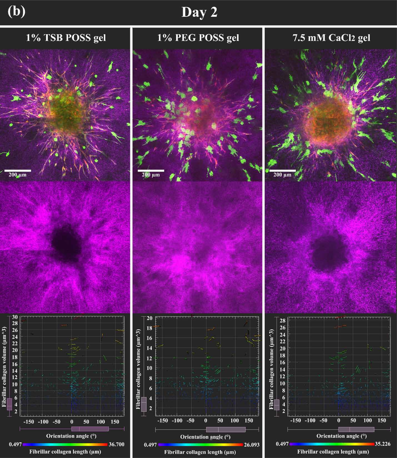
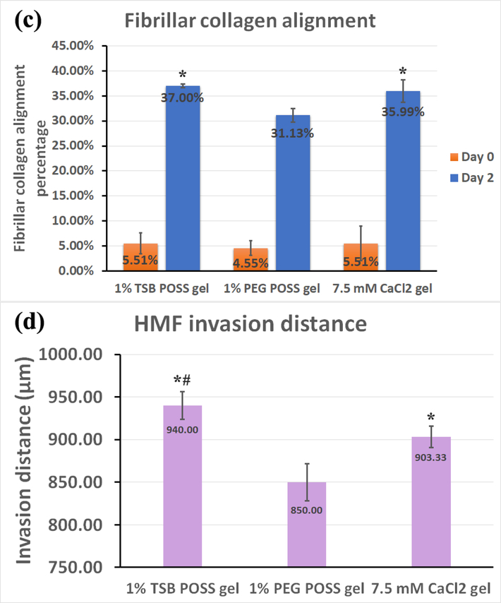
Figure 6. HMF spheroids remodel collagen alignment to regulate cancer cell migration in POSS nanocomposite hydrogels.
We embedded HMF spheroids (shown as overlay of red and green fluorescence) into hydrogels with dissociated MDA-MB-231 cells (green fluorescence). Images show the z-intensity projection of HMF spheroids and cancer cells overlaid on second harmonic imaging of fibrillar collagen matrix (pseudocolor in purple) (top row). (a) On day 0 (2 hours after embedding), HMF spheroids showed no protrusions, and MDA-MB-231 cells retained round morphology without localization to spheroids. Bottom row displays measured orientations of fibrillary collagen, where 0° indicates fibers oriented perpendicular to the spheroid surface (radical alignment). Pseudocolor side bar depicts fiber length. (b) On day 2 (48 hours after embedding), HMFs migrated extensively out of spheroids in a sun-burst pattern. MDA-MB 231 cells showed elongated morphology and migrated towards spheroids (top row). HMFs remodeled the collagen matrix, evidenced by thicker bundles of fibers with alignment coincident with HMF migration pathways (middle row). Distribution of fibrillar collagen orientation angles showed a peak at 0°, indicating more fibers with perpendicular alignment (bottom row). (c) Graph compares alignment of collagen fibers perpendicular to spheroid surfaces in different hydrogels, on days 0 and 2. *: > 1% PEG POSS gel, p < 0.05. (d) Quantification of invasion distances of HMF spheroids in different gels on day 2. *: > 1% PEG POSS gel, p < 0.05; #: > 7.5 mM CaCl2 gel, p < 0.05.


