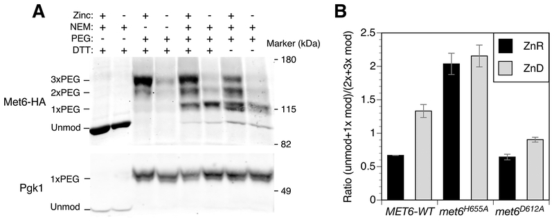
Figure 10.
In vivo analysis of zinc binding by Met6. A) NEM/PEG-maleimide analysis of wild-type (BY4742) cells expressing Met6-HA. Zinc-replete (+) or deficient (–) cells were treated with and without NEM, proteins harvested, and then treated with and without DTT prior to PEG-maleimide treatment. The positions of molecular mass markers (kDa) and unmodified and PEG-modified forms of Met6-HA are shown. Pgk1 was detected as a control for PEG labeling efficiency. B) Quantification of NEM/PEG-maleimide analysis of wild-type (BY4742) cells expressing Met6-HA, Met6H655A-HA or Met6D612A-HA as described for panel A. The ratios of unmodified + 1 × PEG-modified to 2–3 × PEG-modified forms are shown and the error bars indicate ± 1 S.D (n = 3).

