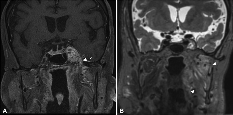
Fig. 2.

Postoperative coronal T1-weighted image with fat saturation ( A ) demonstrates typical imaging characteristics of HB with serpentine flow voids. The mass incompletely encases and medially displaces the cavernous portion of the left internal carotid artery ( A ). The mass exhibits inferolateral extension into the ipsilateral foramen ovale (white arrow; A ) and a nodular projection to the vicinity of the optic tract superiorly. Coronal T2-weighted image ( B ) reveals the sequelae of CN V3 involvement by the tumor with advanced chronic denervation atrophy with fatty replacement of the left medial and lateral pterygoid, masseter and temporalis muscles (white arrowheads; B ). CN, cranial nerve; HB, hemangioblastoma.
