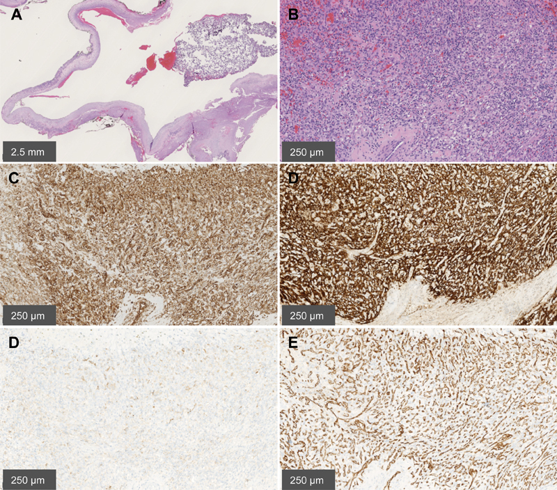
Fig. 3.

Pathologic study of the surgical specimen. ( A ) Low magnification shows a cystic nature of the lesion; haematoxylin and eosin ×10. ( B ) Higher magnification demonstrates the dual component tumor of hemorrhagic, thin-walled, vascular channels lined by flattened endothelium (especially to the left of the picture) and of stromal proliferation with large, polygonal, cells containing vacuolated cytoplasm (especially mid and proximal right of the picture). Note the prominent mast cell infiltrate (small, dark, round nuclei); haematoxylin and eosin ×10. ( C ) Tumor cells are strongly positive for vimentin. Vimentin stain ×100. ( D ) Tumor cells are strongly positive for carbonic anhydrase IX; CA IX stain ×100. ( E ) Tumor cells are focally positive for inhibin; Inhibin stain ×100. ( F ) Vascular channels are highlighted by CD31; CD31 stain ×100. CA, carbonic anhydrase.
