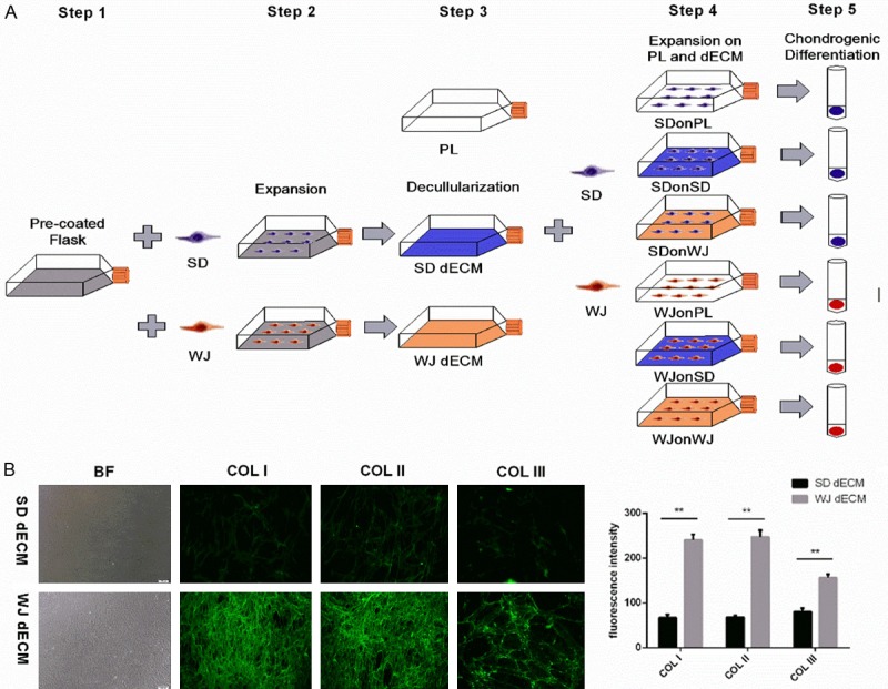
Figure 1.

Decellularized cell matrix (dECM) fabrication and cell expansion and physiochemical properties of dECMs. A. Flow diagram of dECM fabrication and cell expansion. Step 1. surface preparation with 0.2% gelatin. Step 2. synovium-derived stem cell (SDSCs) and Wharton’s jelly-derived stem cell (WJ-MSCs) culture. Step 3. dECM fabrication using SDSCs and WJ-MSCs. Step 4. cell seed and harvest after dECM expansion. Step 5. chondrogenic differentiation. SD: SDSCs, WJ: WJ-MSCs. B. Representative dECMs appearance and immunofluorescence staining for COL I, COL II and COL III in dECMs deposited by SDSCs and WJ-MSCs. Magnification: 100×. ImageJ software was used to quantify the immunofluorescence intensity. The experiments were conducted in triplicate. **P < 0.05.
