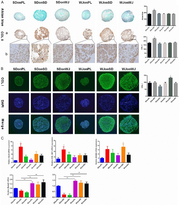
Figure 3.
Evaluation of early (7 days) chondrogenic differentiation. A. Alcian blue and immunostaining were used to detect sulfated glycosaminoglycan (sGAG) and type II collagen deposition of pellets from each group after 7 days of chondrogenic induction and ImageJ software was used to quantify the immunohistological intensity. Magnification: (a) 100× and (b) 200×. The experiments were conducted in triplicate. *P < 0.05 compare with other groups. B. Immunofluorescence staining was used to detect type I collagen deposition after 7 days of chondrogenic induction and 4,6-Diamino-2-phenylindole (DAPI) was used as a nuclear marker. ImageJ software was used to quantify the immunofluorescence intensity. Magnification: 100×. The experiments were conducted in triplicate. *P < 0.05 compare with other groups. C. Real-time polymerase chain reaction (PCR) was used to evaluate chondrogenic marker genes (Sox 9, COL2A1 and ACAN) and hypertrophic marker genes (RUNX2 and MMP13) expression. The experiments were conducted in triplicate. *P < 0.05 compare with other groups, **P < 0.05.

