
Figure 3.
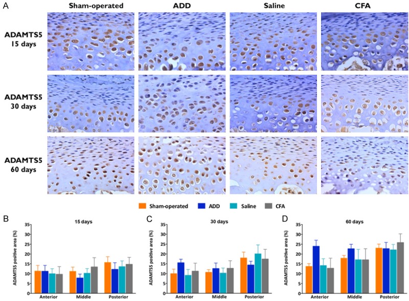
Immunopositivity for the metalloproteinase ADAMTS5. (A) Immunohistochemistry for ADAMTS5 in the TMJ fibrocartilages under x400 magnification. The ADAMTS5 gene was highly expressed in the condylar tissues, mainly in the pre-hypertrophic and in the hypertrophic layers, being only partially expressed in the proliferative layers. Most of the cells of the pre-hypertrophic and the hypertrophic layers were immunopositive for ADAMTS5. (B-D) Marked areas/total area of the ADAMTS5 immunopositive cells in the proliferative, the pre-hypertrophic and the hypertrophic layers, expressed in percentages at 15 (B), 30 (C) and 60 (D) days. No statistical differences were observed, despite that the ADD group showed an increased relative immunopositive area at 30 and 60 days, in their anterior thirds, especially at 60 days. The columns represent the mean of 5-8 independent experiments and the vertical lines indicate the standard error of the mean.