
Figure 4.
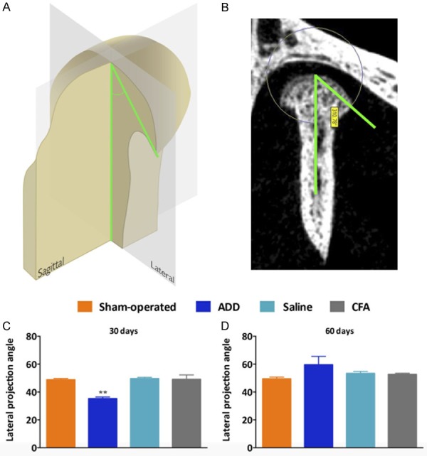
3D reconstruction showing the condylar surfaces and the osteophyte formations. A and B. Representative image and the quantitative analysis of the angle that was formed between the lateral projection of the condyles and the center-coronal axis. C. The ADD group showed a significant reduction of the lateral angle projections when compared with the other groups. All of the samples from the ADD showed the closest angles of projection, associated with the formation of the osteophytes, at 30 days. D. At 60 days, these angles of projection became more open, with no statistical differences among the groups. This was probably correlated with the bone remodeling at the surfaces from the ADD condyles. **P < 0.01 when comparing the ADD to the sham-operated rats.