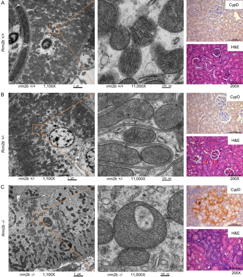
Figure 1.

Rrm2b-knockout mice exhibit mitochondrial swelling and high Cyclophilin D (CypD) expression. Left panel, scanning electron microscope images at 1100-fold magnification (scale bar: 2 µm). A 10 × higher magnification of the area marked by the orange square is located next to the 1100-fold magnification image. Right panel, immunohistochemistry (IHC) stained with anti-CypD (CypD) and hematoxylin and eosin (H&E)-stained images of kidney tissue from Rrm2b+/+ (A), Rrm2b+/- (B), or Rrm2b-/- (C) mice. Scale bar corresponds to 100 µm.
