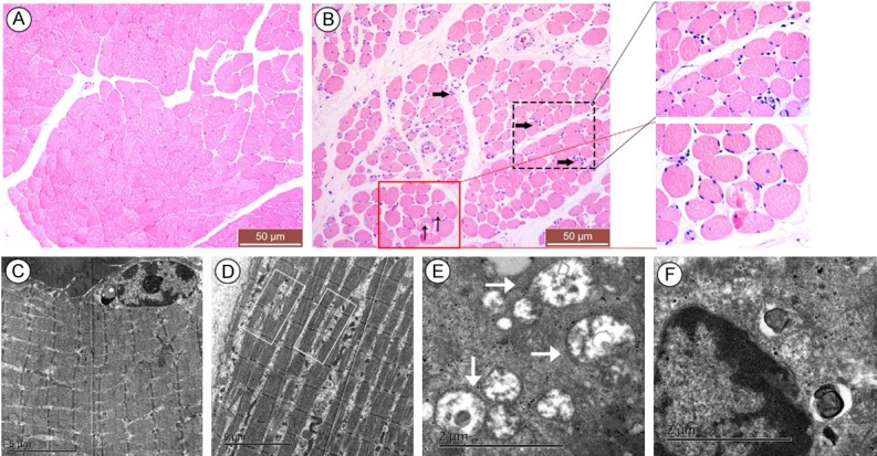
Figure 1.

Histopathology of muscle tissue cells by HE staining and transmission electron microscopy. HE staining of normal muscle tissues showed an angular shape and tightly arranged myofibers with peripheral nuclei (A), while pressure ulcer (PU) muscle tissues demonstrated the morphological characteristics of degeneration, including atrophied or fractured myofibers with a round shape, increased endomysium distance between the fibers, accumulated nuclear aggregation in the interstitial space (as demonstrated by the black thick arrow) and presence of centralized myonuclei (as demonstrated by the black thin arrow); insets are higher magnification (B). However, the ultra-structure of the normal tissue indicated clear internal fine structure and orderly tight arrangement of sarcomeres (C), while PU muscle cells revealed loosely arranged, lysed and discontinued sarcomeres (as demonstrated by the white rectangle in D), swollen denatured mitochondria (as demonstrated by the white thick arrow in E), as well as chromatin margination (F). The scale was shown in corresponding picture.
